 Gregory Albers has been involved in two of the most transformative studies for stroke patient treatment and care in recent times. Here he speaks to NeuroNews about his experiences and how he has seen stroke treatment changes and what he hopes for the future of stroke treatment.
What drew you to medicine and interventional neurology in particular?
My fascination with the brain started in high school. I wanted to understand how memories are made. As an undergraduate, I worked with the late Richard F Thompson who was an expert in learning and memory research. I loved neuroscience, but felt a bit isolated in the lab. I decided to focus on neurology to continue to try and understand the brain as both a doctor and a scientist.
Who were your mentors and what impact have they had on your career?
I began my neurology residency at Stanford University in 1985 and one of my mentors was Dennis Choi, who was studying why brain cells in culture die so quickly when the oxygen and glucose supply is cut off. Dennis was a phenomenal scientist, who was trying to treat stroke in a petri dish with neuroprotective agents. I tried to translate Dennis’ work from the bench to the bedside but this effort was not successful. As a clinical trialist, my mentor was the late David G Sherman who was a pioneer in clinical stroke research. Sherman had a very practical, “keep it focused and easy to execute”, approach to clinical trial design”.
You have been practising for a number of years. How have you seen the field of interventional neuroradiology change and develop over that time?
In the 1990s we were using intra-arterial thrombolytics. They often opened the occluded vessel and we would celebrate in the angiography suite. The next day some patients did great while others developed a huge infarct or brain haemorrhage. We learned that patient selection was going to be the key to successful treatment. We then started doing MRI scans prior to the procedures and were able to determine that it was the patients with the small DWI lesions that were doing great after reperfusion, while the patients who had large DWI lesions were having poor outcomes. Now, 20 years later, we have finally been able to prove in randomized trials that imaging-based patient selection can identify patients who benefit from thrombectomy up to 24 hours from symptom onset.
In your opinion, what has been the most practice-changing advance in terms of treatment options and devices?
The stent-retrievers are primarily responsible for the success of early window thrombectomy that dramatically changed the field in 2015. Now in 2018, it is advanced imaging and automated image processing that are key to extending the benefits of thrombectomy into late time windows. About a decade ago we developed the RAPID imaging software platform for stroke patients at Stanford. Now, following Food and Drug Administration approval and multiple successful clinical trials, the software is being used in more than 350 stroke centres worldwide, both clinically and in ongoing research studies. The RAPID imaging maps show tissue that is likely to be irreversibly damaged and estimate the size of the stroke if reperfusion does not occur. RAPID was used to select patients in SWIFT PRIME and EXTEND IA which were the early window thrombectomy trials that had the largest treatment effects and the highest rates of good outcome. Recently, RAPID was used to select all of the patients in both DAWN and DEFUSE 3 which led to quadrupling the treatment window for thrombectomy from six to 24 hours.
What has been the biggest disappointment?
When I started as a brand-new Assistant Professor in 1989, I thought neuroprotective agents would be the first successful stroke therapy. These agents may have failed because we did not have a way to reliably reperfuse the brain. Eventually collaterals fail, and neuroprotective agents are likely to be successful only if reperfusion occurs. Now that we can identify patients with salvageable tissue and reperfuse them effectively, I think neuroprotective agents need to be reassessed. They could be given during transfer from a primary to comprehensive centres to maintain the penumbra during transport.
What is on your wish list in terms of the future development of stroke therapy?
- Late window intravenous thrombolysis for patients with good collaterals and slow growing infarct cores.
- Thrombectomy for patients with a large ischemic core lesions that are accompanied by a large volume of salvageable penumbra.
- Therapies to improve collateral flow before a patient has a stroke.
You are the chief investigator of the DEFUSE 3 trial which, along with the DAWN trial, helped to pave the way for an extended treatment window for stroke patients. How does it feel to have had a role in this big change, and what further advancements do you see on the horizon?
It feels amazing. Once we started imaging our stroke patients with serial MRI scans we learned that every stroke evolves in a unique manner and we realised that the textbooks were not correct. It is not “all about time”, it is “all about the evolution of the infarct”. Some patient’s infarcts evolve very slowly, other patients, who look identical in the ER and have the same vascular occlusion, evolve very fast. The great news is that the slower evolving patients are more common than the fast growers. This concept has been very unpopular over years because it challenged conventional wisdom. This made it more difficult to get grants funded or papers published. It is incredibly satisfying to see a 20-year-old hypothesis finally changing the field. People are looking at stroke more optimistically now. If thrombectomy can work in extended time windows, why not try late-window intravenous thrombolysis or neuroprotective strategies? Our goal is to have an effective therapy to offer every stroke patient and that therapy should be chosen based on the pathophysiology of the individual patient’s stroke at the time they present for care. Patient selection had a big role to play in the success of those trials.
How was the protocol defined and do you believe that this should be used to select all thrombectomy-eligible patients going forward? Are other selection criteria/modes still viable?
For patients who present ultra-early with a large vessel occlusion to an endovascular centre, virtually all should be treated with thrombectomy. But as the hours start to pass, you begin to see patients with completed infarcts and very large infarct core volumes. Imaging can identify these patients who are more likely to benefit from a therapy to prevent malignant oedema, rather than reperfusion. For later window therapy, advanced imaging is mandatory. All of the “time is brain” studies over the recent decades show us that good outcome rates decline rapidly in unselected patients, even if reperfusion occurs. In contrast, well selected patients with substantial volumes of salvageable tissue can maintain stable rates of good outcome following reperfusion over an extended time period.
What was your most memorable case and what did you learn from it?
The first time I saw a DWI [diffusion-weighted imaging] lesion in a stroke patient. I thought to myself “This is the Holy Grail. Now we can see a stroke evolve in real time. This is the key to understanding stroke evolution.”
What advice do you hope your mentees and students will always follow?
Question what you are taught. Where did the evidence come from? How reliable is it? Are the data being evaluated objectively or could bias be influencing the conclusions? Is there a different angle to look at the problem that might lead to an alternative solution?
What are your interests/hobbies outside of medicine?
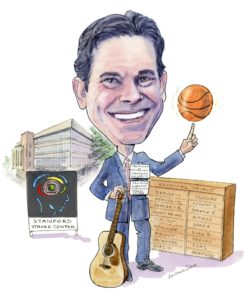
I am a very proud Dad to five very active children. Over the years I have been a coach or spectator at an immense number of baseball, basketball, swimming, water polo, and ballet events. Our family loves to travel, everyone is always up for a new destination. I play the guitar and a few years ago was a member of an all-neurologist group called the Hypertonics. We have performed in San Francisco, Los Angeles and Honolulu, USA.
Fact file
Current research and scholarly interests
- The acute treatment and prevention of cerebrovascular disorders
- Use of advanced imaging techniques to expand the treatment window for ischaemic stroke
- Conducting clinical studies of both neuroprotective and thrombolytic strategies for the treatment of acute stroke
- Investigating new antithrombotic strategies for stroke prevention.
Academic appointments
Professor—Med Center Line, Neurology & Neurological Sciences
Professor—Med Center Line (By courtesy), Neurosurgery
Member, Stanford Neurosciences Institute
Administrative appointments
1992—present Director, Stanford Stroke Center, Stanford Medical Center
2007—present Coyote Foundation Professor, Neurology and Neurological Sciences
Professional education
2008 Board Certification: Vascular Neurology, American Board of Psychiatry and Neurology
1990 Board Certification: Neurology, American Board of Psychiatry and Neurology
1988 Residency: Stanford University School of Medicine Registrar
1985 Internship: Stanford University School of Medicine
1984 Medical Education: University of California San Diego School of Medicine


