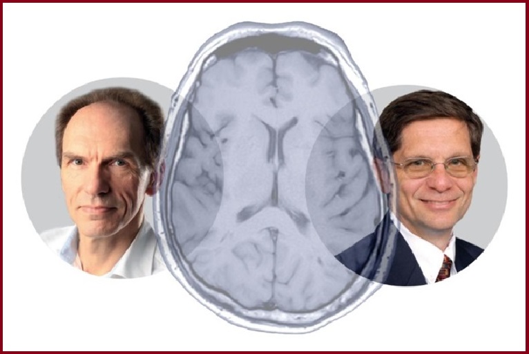
Mechanical thrombectomy has gone from strength to strength in the treatment of acute ischaemic stroke over the past decade—progressing beyond early-window large vessel occlusions (LVOs) to patients presenting >6 hours post-symptom onset and those with occlusions located in the posterior circulation. The latest frontier thrombectomy appears to have conquered is those cases characterised by more extensive infarcts on imaging, generally referred to as ‘large-core’ strokes. However, as alluded to by both Wim van Zwam (Maastricht University Medical Center, Maastricht, The Netherlands) and Joseph Broderick (University of Cincinnati College of Medicine, Cincinnati, USA) in recent interviews with NeuroNews, a number of important questions remain.
Earlier this year, the LASTE trial became the latest addition to an ever-growing list of published randomised controlled trials (RCTs) indicating the safety and clinical benefit of mechanical thrombectomy in ischaemic strokes with large ischaemic cores. Outlining their results in the New England Journal of Medicine (NEJM), the LASTE investigators report that thrombectomy plus medical care resulted in better functional outcomes and lower mortality than medical care alone, but also led to a higher incidence of symptomatic intracerebral haemorrhage (sICH), across a population of acute stroke patients with a large infarct of unrestricted size presenting within seven hours of symptom onset.
The past three years have seen a number of similar RCTs presented to the world, starting with the Japan-based RESCUE-Japan LIMIT trial in early 2022, which established the notion that thrombectomy may be safe and more effective than the existing standard of care for these large-core stroke patients. This has since been followed by the Chinese ANGEL-ASPECT and global SELECT2 trials, and the largely European TENSION study, all of which met their primary endpoint and echoed prior indications of the benefits to treating large-core patients endovascularly. TESLA—a solely USA-based RCT—very marginally missed its three-month primary endpoint for demonstrating thrombectomy’s effectiveness versus medical care, but generated a more successful finding at one-year follow-up.
While these studies are being grouped together as ‘the large-core trials’, they all differ from one another in a handful of subtle yet meaningful ways. Notably, where RESCUE-Japan LIMIT, ANGEL-ASPECT and SELECT2 all primarily defined a ‘large core’ as one characterised by an Alberta stroke programme early computed tomographic score (ASPECTS) of 3–5, TESLA included a wider range of core volumes (ASPECTS 2–5). In addition, a point that TESLA and TENSION have prided themselves on is their use of more pragmatic imaging protocols, solely utilising non-contrast CT scans to define core volumes. ANGEL-ASPECT and SELECT2, on the other hand, have integrated CT perfusion or diffusion-weighted magnetic resonance imaging (DW-MRI) into their assessments of infarct sizes. And, continuing this trend, ASPECTS ranges and imaging protocols are among the minutiae upon which LASTE appears to have broken new ground.
“A unique feature of our trial was the lack of restriction on the upper limit of the infarct size,” the LASTE authors write. “Consequently, 56% of the patients in our trial had a baseline infarct size (ASPECTS value ≤2) that would have precluded their enrolment in other trials that included patients with a large core. Furthermore, the median baseline infarct volume of 135ml in our trial was larger than that in other trials, which may explain why the percentages of patients who died or had severe disability were higher than those in other trials. Nonetheless, the effect favouring thrombectomy was similar in magnitude to that seen in other thrombectomy trials, including those that only enrolled patients with a small or moderately sized baseline infarct; however, no direct comparisons with other trials can be made because of differences in trial designs and patient populations.”
TESLA in context
One point that Van Zwam is keen to address when homing in on the results of these studies is the perception of TESLA and its findings; having missed its three-month primary endpoint, the trial is more likely than any of its large-core contemporaries to be thought of as a ‘negative’ research endeavour. However, Van Zwam points out that a key driver of this outcome was the triallists’ use of Bayesian statistical analyses—as opposed to a more conventional approach, which would have seen the trial reach its primary endpoint and likely come to be viewed in an even more positive light.
“They had to present it as a ‘neutral’ trial that missed the threshold of its primary endpoint,” Van Zwam comments. “But, for me, it’s a positive trial. If you compare it with the other trials, the treatment effect is just as large, so it’s a matter of how you interpret the data.”
As mentioned previously, the TESLA trial also came to reach its primary success measure upon evaluation of the 12-month follow-up data, and has since been joined by both SELECT2 and TENSION in achieving positive results in the long term. The apparent uptick in thrombectomy’s clinical benefits from three months to one year led TESLA’s presenting authors to conclude that—in addition to highlighting the importance of continued follow-up and rehabilitation—their findings point to the better recovery and overall improvement in functional outcome enabled by thrombectomy. Van Zwam agrees with this conclusion, adding that the observed effect is demonstrative of the fact that, over a longer period of time, patients in control groups receiving only standard medical care without thrombectomy do even worse versus those who are treated interventionally, resulting in a more noticeable divergence in functional outcomes between the two.
“It’s something that we’ve also seen with the MR CLEAN-LATE trial. We didn’t see a difference in mortality [between thrombectomy and controls] after three months, but there was a difference after two years,” he continues, stating that a similar trend appears to be present in patients with large-core strokes—a group that, in general, already experiences worse outcomes versus those with smaller infarcts. “That is the mechanism we see with longer follow-up. We saw it in REVASCAT as well; after one year, the treatment benefit is still there, and mortality is increasing in the control group. And it makes sense that the treatment effect in larger cores will be even stronger after a longer period, because the control groups in these low-ASPECTS trials do very badly.”
Truly defining ‘large core’
According to Van Zwam, the fact that TESLA initially missed its primary endpoint makes the results of LASTE “even more spectacular, because around 50% of its patients had an ASPECTS of 0–3, with the other half being ASPECTS 4–5”.
“In LASTE, we were afraid that those very severe, very low-ASPECTS patients would dilute the effect of thrombectomy and it would not produce significantly positive results—but it was positive [at the six-month follow-up], even though it had that large cohort with very low ASPECTS,” he explains. While LASTE has been lauded as the latest in a long line of momentous breakthroughs with stroke thrombectomy, the neurointerventional community is still faced with a handful of unresolved challenges. And, in Broderick’s view, despite the overwhelming positivity surrounding them, the published trials in this space do carry their own limitations—with one of these being a degree of “overlap” in how each of them defines large core.
“First of all, I think almost all of them started with patients who really had no disability or minimal disability to begin with, meaning they’re a very pristine group—they’re not people who already have some functional disabilities, brain injuries or a lot of prior strokes. So, that’s one thing you’re taking out,” Broderick states. “In addition, I think our data on large-core patients with ASPECTS ≥3 are pretty good, but data on ASPECTS 0–2 are still very limited. And, also, when you get to large cores as defined by perfusion studies, we don’t have a lot of data beyond the 100cc range. We also don’t have a lot of data on the very old. So, there are a lot of questions that still have to be answered.”
Van Zwam and Broderick are in agreement about another area requiring further clarity: potential discrepancies between a patient’s ASPECTS and the true ‘core size’ of an ischaemic stroke. While a low ASPECTS has become something of a proxy for general stroke severity, Van Zwam notes that ASPECTS primarily indicates early signs of ischaemia and reduced perfusion, but does not provide conclusive information on the extent to which areas of brain tissue are already ‘dead’ or unsalvageable. This is because some of the damage may in fact be reversible if the affected blood vessel can be reopened and reperfused once more. According to Van Zwam, this notion has been confirmed via studies looking at final infarcts in which stroke severity has turned out to be lower than anticipated based on ASPECTS.
“There is a lot of research in progress right now, and these trials help enormously, but a single item like large core on perfusion CT or like ASPECTS probably does not define whether a patient should receive thrombectomy or not, or their chance of a good outcome,” Van Zwam explains, also highlighting areas of demarcated hypodensity on imaging as potentially being a better indicator of ‘real core’ that is truly unsalvageable. However, even this metric is unable to account for the convergence of numerous variables—brain frailty, old age, atrophy, hyperglycaemia and hypertension as well as time last known well—that all play into thrombectomy-related decision-making.
“We should, in fact, be very liberal in treating patients with all of those early signs of infarction because, even in the late window, those signs don’t define anything beyond the severity of the symptoms when the patient enters the hospital. ASPECTS shows the area that is affected but it doesn’t tell us what tissue is still salvageable. I think, when you have ASPECTS 1–2—especially in the very early window—the patient can still do very well if you treat them [with thrombectomy] very quickly. In the later window, you may have to be a bit more selective, but we still suspect that ASPECTS is not a good discriminator between who will benefit from treatment and who will not.”
In addition, the early signs of ischaemia on imaging are often very subtle, with several factors contributing to their appearance, meaning the determination of ASPECTS can also be highly subjective.
“There are whole discussions going on right now regarding those baseline CT scans,” Broderick adds. “How dark is dark on ASPECTS? What’s the measurable water content in the brain? One term I’ve heard used recently is that, while there’s the ‘dark brain’ that’s pretty much dead, maybe there’s the ‘dusty brain’ that might not be dead yet but is getting close. So, again, there’s a lot of variability in definitions and a lot of variability in the populations being studied, and it’s going to be important for us to work out—with these larger areas of damage—who best qualifies for treatment, as well as being realistic about the benefit itself.”
The timing of ASPECTS measurement is also thought to hold some relevance, as earlier imaging—either on CT or MRI—is a less clear indicator of ‘dead brain’ as compared to imaging performed in the later time window. On this point, Broderick highlights the fact that LASTE required ASPECTS imaging to be done within 6.5 hours.
Another point he makes is that determining ASPECTS based on CT imaging or MRI is not the only way that a large core can be defined. An alternative approach is via diffusion-weighted changes, which Broderick says gives a clearer indication of tissue that is likely to be unsalvageable versus parts of the brain that are not yet ‘dead’ despite appearing to have diminished blood flow. However, this discussion is something of a balancing act because, while DW-MRI—as compared to baseline CTs—is likely to be a more accurate and less ambiguous indicator of irreversible brain damage, CT imaging offers a significantly more pragmatic and easy-to-use solution that is far more widely available and utilised at a greater proportion of centres.
“I don’t think MRI is going to end up being the standard imaging tool for stroke patients in the emergency department, at least for a while,” Broderick speculates. “It is in some places, where they have it set up and they have very good programmes—but CT is just so fast, and it provides a lot of information to help make initial decisions, so I don’t see its role being usurped by MRI at this moment in time.
“The other thing is that, in some places, they go right to the angio suite and do a ‘poor man’s’ CT—but a reasonable CT—with the equipment they have there. Again, this saves time, and time really is brain. It’s been a frequent refrain for many, many years, but it’s true, so anything that can get you the key information that you need to help make the best decisions is going to be the predominant way that we treat patients.”
New frontiers
Looking back to 2013, when three RCTs—IMS III, MR RESCUE and SYNTHESIS—were published, and suggested unanimously that thrombectomy should not be considered an appropriate treatment for LVO acute ischaemic stroke, the situation stands in stark contrast to the groundswell of enthusiasm surrounding the intervention today. Patient inclusion criteria, treatment time windows, imaging protocols and the quality of thrombectomy devices all hampered those earlier studies, before being optimised ahead of the five positive RCTs published just two years later.
It may therefore come as little surprise that greater refinement in each of these areas, in addition to an improved understanding of the mechanisms behind the procedure and ever-growing levels of operator experience, has led to positive results in subsequent trials seeking to expand thrombectomy’s range further still. LASTE and the other large-core RCTs, as well as the 2018 DAWN and DEFUSE-3 studies extending the appropriate therapeutic window to 6–24 hours, and the more recent ATTENTION and BAOCHE trials supporting the endovascular treatment of basilar-artery occlusions (BAOs), are all notable examples. As Van Zwam quips, “nowadays, it seems that whatever trial we do will be positive, because the treatment effect is so significant”. Nonetheless, in the minds of both Van Zwam and Broderick, further studies will add critical information to the neurointerventional community’s current knowledge base. Here, Van Zwam highlights stroke patients with very mild symptoms and low National Institutes of Health stroke scale (NIHSS) scores as a noteworthy area of interest moving forward, with strokes caused by more distal or medium-vessel occlusions (D/MeVOs) being another.
However, on the latter front, he advises a degree of caution, citing the fact that many skilled physicians already routinely operate on more distally located occlusions—and have done for many years following the positive LVO stroke trials of 2015—despite a multitude of RCTs specifically investigating these cases still being in progress right now. A second area where some discrepancy remains is the aforementioned basilar artery: while the successes of the Chinese ATTENTION and BAOCHE alleviated some concerns in this location following more mixed results from BASICS and BEST, one of the drivers of this was their decision to focus solely on BAO patients with more severe deficits.
“We sometimes become overenthusiastic and think that all treatments will be beneficial, which is ‘nearly true’,” Van Zwam opines. “Most of the trials we do now are positive, but there are a few exceptions. I don’t think we should always treat the BAOs with very mild symptoms endovascularly. And the more distal occlusions with mild symptoms—maybe not, but we have to wait for the trial results. Even at conferences, you see that companies often offer dedicated devices for very distal occlusions, and physicians present cases where they go very distal with a tiny device and show that they can open the vessel. But I don’t know if, in general, this is a good thing to do. We have to wait.”
The bottom line
“I think we’re going to find that there are limits to thrombectomy [in the brain], like there are in the heart—they have the same issue with myocardial infarction and we’re about 10 years behind them,” Broderick adds. “We have slightly different situations in terms of what causes the strokes and even the devices we use to treat them. The recent large-core trials are a step forward, and we have a lot of data to work with, but there are still a lot of questions that are unanswered.”
In addition, while LASTE has created an air of excitement around the idea that thrombectomy’s effect may not be curtailed by even the largest infarct sizes, Broderick believes it is important for interventionists to remain realistic and reasonable regarding the benefits they expect to see with endovascular treatment versus best medical care.
“We have to take into account the patient’s functional status and what the family’s wishes are,” he says. “What do you do in the case of somebody who already has a major deficit and experiences a large-core stroke? The best you can hope for is to get them back to the same deficit or, more probably, a less functional state than before.
“I also think that, while there’s been a lot of excitement about large core, there has been some pushback from the interventional community, because people just don’t like to take care of patients who—even if you open up their artery and they do a little bit better—are still going to be pretty affected by their stroke. People prefer to take the patients who are younger and, when you reopen their vessel, they’re able to walk again and talk again. A lot of the more severe large-core stroke patients still die or have a very bad outcome, even though we’ve moved the needle in the right direction with endovascular treatment.”
Broderick comments that it will likely take time for physicians to become more comfortable with the existing data and how they relate to routine practice, and—as he puts it—this continued angst over whether or not to treat certain patients proves that, “even among the interventional community, it’s not all smiles and rainbows”.
“However, I think the main message is that, for patients who appear to have a reasonable amount of brain injury, some of them can still have an [acceptable] outcome—they don’t all return to full functional ability but, if you look at a scale of how well they function, there is some improvement compared to those who did not get any kind of intervention,” he continues. “And the bottom line is that, as long as we’re doing good, randomised trials, and we’re being as inclusive as possible, we’ll gain more and more information about who best qualifies and who doesn’t qualify for thrombectomy. It’s about getting the best data from the scientific trials and then applying them to the individual situation, and the individual patient.”
To conclude, Broderick draws attention to an upcoming statement being worked on by the American Heart Association (AHA) that will “provide a lot more clarity” when it comes to the large-core trials—adding that, “while we’ve made a lot of progress, that statement will really help to illustrate what we know and what we don’t know”.


