Two analyses disclosed for the first time at the 11th European Stroke Organisation Conference (ESOC; 21–23 May, Helsinki, Finland) have shed fresh light on the potential role for mechanical thrombectomy in acute ischaemic strokes caused by distal/medium-vessel occlusions (D/MeVOs), following presentations of neutral efficacy results across three randomised trials comparing the procedure to best medical therapy earlier this year.
The first of these analyses saw investigators conduct a sub-study of the DISTAL randomised controlled trial (RCT), ultimately revealing that the addition of endovascular thrombectomy to best medical therapy led to an increased probability of brain tissue being preserved in D/MeVO stroke patients.
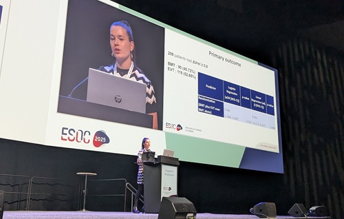
“This study gives a different perspective into how we interpret treatment success in D/MeVO stroke,” said Aikaterini Anastasiou (University Hospital Basel, Basel, Switzerland), who presented these results at ESOC 2025. “Rather than focusing solely on a functional score that potentially does not capture improvements of less severe strokes, we could also consider the extent of brain tissue that can be saved.”
The main results of the DISTAL trial showed no improvement in functional outcomes with thrombectomy over best medical therapy in D/MeVO stroke patients at 90 days—a phenomenon that the researchers believe could be explained by the smaller tissue territory at risk and lower National Institutes of Health stroke scale (NIHSS) scores in these cases as well as greater procedural risks potentially offsetting the benefits of reperfusion. These data, along with findings from the ESCAPE-MeVO and DISCOUNT RCTs, have sparked debate over thrombectomy’s true role in stroke patients with more distal occlusions.
By focusing on the amount of ‘threatened’ brain tissue that was actually saved with the treatment, as opposed to the more traditional clinical endpoint of functional independence on the modified Rankin scale (mRS), Anastasiou and colleagues’ new imaging-based analysis attempted to approach the matter from a different perspective.
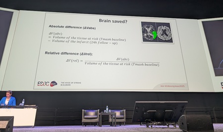
Using baseline perfusion and 24-hour follow-up imaging from 447 trial participants, they assessed the proportion of brain tissue at risk of infarction that was ultimately spared, and found that patients receiving thrombectomy plus best medical therapy had a higher likelihood of achieving a good imaging outcome—having at least 80% of their at-risk tissue preserved—compared to those receiving best medical therapy alone (53% vs 41%). This sub-analysis showed that saving more than 80% of a patient’s threatened tissue was also found to be strongly associated with better functional outcomes at discharge and at 90 days in both treatment groups.
“These results suggest that, even in patients with smaller strokes due to D/MeVO, the amount of salvaged brain tissue plays a critical role in recovery,” Anastasiou added. “Our analysis indicates that successful reperfusion through [endovascular thrombectomy] can significantly increase the likelihood of saving brain tissue—and, in turn, improve outcomes.”
Another key finding was the fact that patients with successful reperfusion, defined by a modified thrombolysis in cerebral infarction (mTICI) score of ≥2b, had an even greater likelihood of significant tissue salvage, as indicated by an adjusted odds ratio (OR) of 2.54. The analysis also found no difference in outcomes between groups when less than 80% of the tissue was saved, which the researchers feel underscores the importance of achieving high-quality reperfusion.
A closer look at ESCAPE-MeVO
A second analysis—presented in the same late-breaking data session at ESOC 2025—shone a spotlight on more granular findings from the ESCAPE-MeVO RCT, in which primary endpoint findings on thrombectomy versus best medical therapy were similarly neutral.
Delivered by Michael Hill on behalf of his co-principal investigator Mayank Goyal (both University of Calgary, Calgary, Canada), and their ESCAPE-MeVO collaborators, this post-hoc imaging analysis revealed a potential link between a D/MeVO stroke patient’s collateral perfusion status on computed tomography (CT) angiography and their eventual clinical outcome.
“These data suggest that, among patients with stroke due to D/MeVO with very poor or absent collateral filling on baseline CT angiography, [endovascular thrombectomy] may be harmful—and, in those with excellent collaterals, [endovascular thrombectomy] may be beneficial,” Hill commented. “We hope to confirm these observations in future pooled analyses of these trial data.”
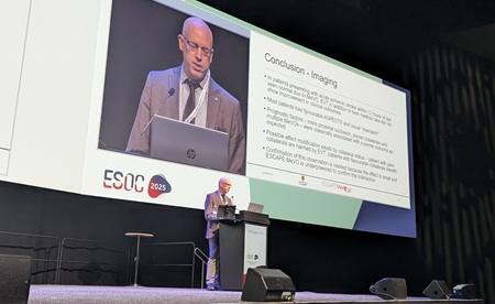
Based on this analysis, collateral status was deemed to be the “only” imaging feature significantly associated with 90-day functional outcomes in D/MeVO stroke patients, as factors including occlusion location and vessel diameter did not appear to have tangibly modified the effect of thrombectomy. These findings may help refine selection criteria for reperfusion therapies in D/MeVO stroke, according to the investigators.
Hill and colleagues’ goal with this analysis was to identify whether these or any other specific baseline imaging characteristics were associated with prognosis, and with the efficacy of intravenous thrombolysis (IVT) and endovascular thrombectomy, in the ESCAPE-MeVO trial. All 529 patients from the trial’s intention-to-treat population were included, with CT angiography being used to determine occlusion location, and to grade collateral status as good, moderate or poor. Univariable and multivariable ordinal logistic regression models were used to evaluate associations of imaging variables with the primary endpoint of 90-day mRS, and with treatment effects of IVT and thrombectomy.
Some 84.7% of MeVOs included in the trial involved the middle cerebral artery (MCA) territory, with M3 being the most common segment (41.4%), while the median occlusion diameter was 1.9mm. Good, moderate and poor collateral statuses on CT angiography were observed at rates of 24.4%, 52.2%, and 21%, respectively.
Regarding prognostic associations on univariable analyses, the researchers observed that each one-tier worsening in collateral grade was linked to a 25% reduction in the odds of a favourable outcome being achieved (common OR, 0.75)—a finding that remained ‘directionally consistent’ upon multivariable analyses (adjusted OR, 0.82). In Hill’s view, this goes some way to confirming the notion that, the more distal a patient’s occlusion is, the less relevant their collaterals are likely to be in terms of impacting post-thrombectomy outcomes.
The ESCAPE-MeVO investigators believe that, based on these results, CT angiography-based collateral status has now emerged as a potential predictor of functional outcomes after D/MeVO stroke, and may inform patient selection for reperfusion strategies, with further studies set to help stratify patients who are more likely to benefit from interventional treatment.
“Time is brain—again”
During his presentation of this ESCAPE-MeVO sub-analysis, Hill drew attention to another potential modifier of the effect of thrombectomy treatment that was apparent in their results: speed.
In addition to the presence of ‘poor’ collaterals, a greater period of time between stroke symptom onset and thrombectomy treatment was linked to worse functional outcomes following the procedure. According to Hill, thrombectomy patients treated within three hours did “much better” clinically, while there was a trend towards thrombectomy being “harmful” as compared to best medical therapy in patients treated 3–12 hours post-stroke. He described this relationship between time to treatment and outcomes as a “qualitative and quantitative interaction” in their analysis.

“Treat early and treat fast” was among the subsequent messages imparted by Hill during his ESOC presentation, as was the assertion that—in contrast to large vessel occlusion, where there is a long tail of patients who benefit from thrombectomy in later windows—“we have neglected speed” in recent times.
“We do have to re-emphasise the need for speed, and we will need to confirm these results in the other MeVO studies,” he added, highlighting time to treatment as one of the few factors that appeared to have a genuine treatment-modifying effect in ESCAPE-MeVO.
These suggestions drew a mixed response from those in attendance at ESOC; DISTAL trial co-principal investigator Marios Psychogios (University Hospital Basel, Basel, Switzerland) commented that the same time-outcome interaction was not observed in his team’s research, while session moderator Mira Katan—also of University Hospital Basel—posited that “time is brain, again”.


