In what has been described as the first study of its kind, researchers from the Keck School of Medicine at the University of Southern California (USC) in Los Angeles, USA and the California Institute of Technology (Caltech) recently designed and implanted a ‘transparent window’ in the skull of a patient, before using functional ultrasound imaging (fUSI) to collect high-resolution brain imaging data through the window.
Their preliminary findings suggest that this sensitive, non-invasive approach could open new avenues for patient monitoring and clinical research, as well as broader studies of how the brain functions.
“This is the first time anyone had applied functional ultrasound imaging through a skull replacement in an awake, behaving human performing a task,” said Charles Liu (USC Neurorestoration Center, Los Angeles, USA). “The ability to extract this type of information non-invasively through a window is pretty significant, particularly since many of the patients who require skull repair have or will develop neurological disabilities. In addition, windows can be surgically implanted in patients with intact skulls if functional information can help with diagnosis and treatment.”
The research participant—a 39-year-old patient named Jared Hager—sustained a traumatic brain injury (TBI) from a skateboarding accident in 2019. During emergency surgery, half of Hager’s skull was removed to relieve pressure on his brain, leaving part of his brain covered only with skin and connective tissue. Because of the COVID-19 pandemic, he had to wait more than two years to have his skull restored with a prosthesis.
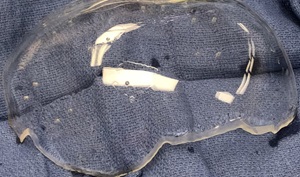
During that time, Hager volunteered for earlier research conducted by Liu, Jonathan Russin (USC Neurorestoration Center, Los Angeles, USA) and another Caltech team on a new type of brain imaging called functional photoacoustic computerised tomography (fPACT). The experimental technique had been performed on soft tissue, but could only be tested on the brain in patients like Hager who were missing a part of their skull. When the time came for implanting the prosthesis, Hager again volunteered to team up with Liu and his colleagues, who designed a custom skull implant to study the utility of fUSI—which cannot be done through the skull or a traditional implant—while repairing Hager’s injury.
Before the reconstructive surgery, the research team tested and optimised fUSI parameters for brain imaging, using both a phantom and animal models. They then collected fUSI data from Hager while he completed several tasks, both before his surgery and after the clear implant was installed, finding that the window offered an effective way to measure brain activity. The research, funded in part by the National Institutes of Health (NIH), has been published in the journal Science Translational Medicine.
As stated in a recent press release from the Keck School of Medicine, fUSI may eventually offer a “sensitive and precise” alternative to current methods for functional brain imaging like functional magnetic resonance imaging (fMRI) or intracranial electroencephalography (EEG), where challenges include low resolution, a lack of portability, and the need for invasive brain surgery.
“If we can extract functional information through a patient’s skull implant, that could allow us to provide treatment more safely and proactively, including to TBI patients who suffer from epilepsy, dementia or psychiatric problems,” Liu added.
In addition to better monitoring of patients, the new technique could offer population-level insights about TBI and other neurological conditions. It could also enable scientists to collect data on the healthy brain and learn more about how it controls cognitive, sensory, motor and autonomic functions, the release adds.
“What our findings show is that we can extract useful functional information with this method,” Liu continued. “The next step is: what specific functional information do we want, and what can we use it for?”
Until the new technologies undergo clinical trials, fUSI and the clear implant are experimental. In the meantime, the research team is working to improve its fUSI protocols to further enhance image resolution. Future research should also build on this early proof-of-concept study by testing more participants to better establish the link between fUSI data and specific brain functions, according to the researchers.


