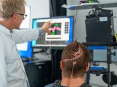
 Inbrain Neuroelectronics has announced the interim analysis of findings from the “world’s first” in-human clinical study of the company’s graphene-based brain-computer interface (BCI) technology. The study—sponsored by the University of Manchester and conducted at the Manchester Centre for Clinical Neurosciences in Manchester, UK—is evaluating the safety and functional performance of graphene-based electrodes when used during the surgical resection of brain tumours.
A press release from Inbrain states that the primary objective of the study is to assess the safety of the company’s BCI during brain tumour surgery, while secondary objectives include evaluating the quality of neural signals captured by the device, its ability to deliver targeted brain stimulation, the consistency of its performance throughout the procedure, and its overall suitability for use in the neurosurgical operating room.
A total of 8–10 patients are expected to be enrolled to validate the safety and functional performance of the graphene-based BCI. The study design included an interim analysis after the first four patients had been recruited to ensure patient safety and data quality.
According to Inbrain, this interim analysis of results from the first cohort of enrolled patients has demonstrated no device-related adverse events—a key component of the primary endpoint of the study. During awake language mapping, the device captured distinct high gamma activity linked to different phonemes—the smallest units of sound in speech—showcasing “exceptional” spatial and temporal resolution, even with micrometre-scale contacts. The ultra-thin, sub-micrometre graphene electrodes also proved compatible with commercially available, CE-marked electrophysiology systems, reliably recording real-time brain signals throughout the surgical procedures, Inbrain claims.
“The ability to detect high-frequency neural activity with micrometre-scale precision opens new possibilities for understanding brain tumour interactions and broader brain function in neuro-related disorders,” said David Coope (Manchester Centre for Clinical Neurosciences, Manchester, UK), the study’s chief investigator. “This technology could be transformative, not only for improving surgical outcomes but for unlocking new treatment pathways.”
Inbrain says in its recent release that, throughout the procedures, its BCI enabled high-resolution brain signal monitoring, addressing “one of the most pressing challenges” in neurosurgery: achieving precise tumour removal while preserving essential functions like speech, movement, and cognition. The device was used in parallel with standard clinical monitoring tools, maintaining consistent performance across the surgical window.
“This milestone demonstrates that graphene-based BCIs can be deployed in the operating room and deliver a level of neural fidelity not achievable with traditional materials,” said Carolina Aguilar, chief executive officer (CEO) and co-founder of Inbrain. “We’re moving toward a future where neurosurgeons and neurologists can rely on real-time, high-definition brain data to guide personalised interventions.”
The release goes on to detail that Inbrain’s platform is powered by ultra-flexible, thin-film graphene semiconductors that conform more precisely to the brain’s surface than conventional strip electrodes. The BCI features high-density, multiscale, bidirectional contacts for superior decoding and modulation, and reduced graphene oxide (rGO) nanoporous matrices that enhance sensitivity and signal resolution. In preclinical studies, the BCI significantly outperformed platinum-based electrodes in detecting high gamma frequencies (80–130Hz) that are “critical” for speech decoding, with statistical significance (p<0.01).
Regarding the potential clinical advantages of its BCI solution, Inbrain claims that graphene technology offers several benefits for neurosurgical procedures. It enhances surgical precision by enabling smaller and more densely packed electrodes, allowing surgeons to define and preserve critical functional areas during tumour resection. Its flexibility enables accurate decoding and mapping in anatomically complex or hard-to-access brain regions, including the walls of the tumour resection cavity. Additionally, the device’s ability to decode high-frequency activity offers “huge scientific opportunities”, including the potential to reveal in situ interactions between glioma cells and neurons, offering potential insights into new therapeutic targets for halting tumour progression.
“We’re not delivering incremental innovation, we’re enabling entirely new capabilities,” said Kostas Kostarelos (University of Manchester, Manchester, UK), co-founder of Inbrain and chief scientific investigator of the study. “This convergence of advanced materials science, neuroscience and AI [artificial intelligence] is shaping the future of real-time, precision neurology.”


