 Royal Philips has today announced the results of a health economics analysis published in the Journal of NeuroInterventional Surgery (JNIS) that show an “innovative approach” to the stroke care pathway—referred to as direct-to-angio suite (DTAS)—can reduce costs by an average of €2,848 (~US$3,120) per patient.
The retrospective analysis looked at data from the controlled, single-centre ANGIOCAT clinical trial conducted at the Vall d’Hebron University Hospital Stroke Unit (Barcelona, Spain). Earlier results from this study have demonstrated that a DTAS pathway is able to improve clinical outcomes for patients who have suffered a stroke.
“The ANGIOCAT clinical study has already shown that bringing stroke patients directly to the angio suite improves patient outcomes,” said Manuel Requena (University Hospital Vall d’Hebron, Barcelona, Spain). “The economic analysis of the data now tells us we can also significantly reduce costs. This indicates that the initial up-front investment of a DTAS workflow will result in a fast return on investment for healthcare providers.”
As a press release from Philips details, after initial triage in the emergency department, the typical treatment pathway for stroke involves sending the patient to the radiology department for a diagnostic computed tomography (CT) or magnetic resonance imaging (MRI) brain scan. This adds time, often worsened by gaps in communication, information and access to stroke expertise.
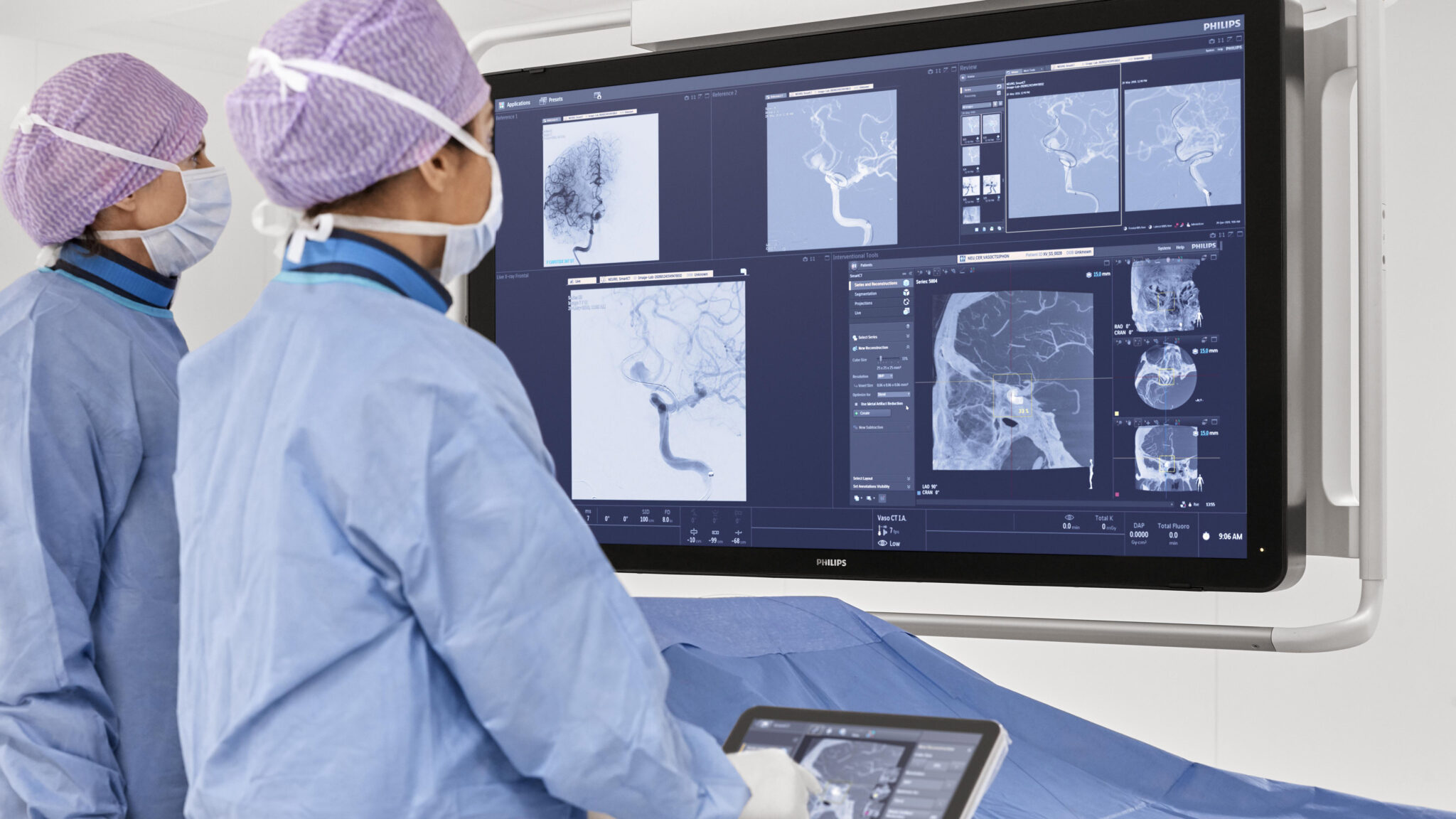
For stroke centres, a time-saving alternative is to have a dedicated angio suite permanently on standby to which stroke patients can be transferred immediately after admission. Using cone-beam CT imaging—such as that built into Philips’ image-guided therapy system, Azurion—clinicians can make a diagnosis and intervene on the spot, saving precious time.
The health economics analysis published in JNIS indicates that a positive return on investing in a dedicated angio suite can be achieved in only a few years, according to Philips.
The company’s DTAS workflow is enabled by an advanced cone-beam CT brain scan performed directly in the angio suite to diagnose patients. Cone-beam CT utilises a cone-shaped beam of X-rays and a flat-panel detector mounted on a C-arm gantry similar to that routinely used in an angio suite, capturing multiple images from different angles to reconstruct 3D images of the brain.
Thanks to technological breakthroughs, Philips claims to have increased the diagnostic confidence of cone-beam CT from 32% to 93% over the course of the past few years. This technology can rule out intracranial haemorrhages (ICHs) and identify large vessel occlusions (LVOs), which account for roughly 25–50% of all acute ischaemic strokes. Patients diagnosed with an LVO can then be immediately operated on using a minimally invasive, image-guided procedure known as a mechanical thrombectomy.
Multiple single-centre studies have shown the positive impact of DTAS on clinical outcomes, resulting in many dedicated stroke centres having already adopted it. A large, multicentre, randomised clinical trial called WE-TRUST is currently running to confirm the patient benefit of DTAS as well.


