 The findings of the first clinical study to use the EVIAS (Biomodex) 3D printing models to plan aneurysm treatment using endovascular robotics and novel flow diverters have been published in the Journal of NeuroInterventional Surgery (JNIS).
The authors, Vitor Nagai Yamaki of Universidade de São Paulo (São Paulo, Brazil) and colleagues concluded that pre-procedural rehearsal using patient-specific 3D models provides precise procedure planning, which they say “can potentially lead to greater operator confidence, decreased radiation dose and improvements in patient safety, particularly in first in-human experiences”.
The study included eight patients with their respective EVIAS 3D aneurysm models. Pre-operative simulation was performed for the first in-human robotic-assisted neurovascular interventions (n=2) and new generation flow-diverter stents (n=6). According to the authors, aneurysms were located in both the anterior (n=5) and posterior (n=3) circulation and were on average 11.0±6.5mm in size.
“We found reliable reproduction of the aneurysm features and similar dimensions of the parent vessel anatomy between the 3D models and patient anatomy,” Yamaki et al write. The team also state that information learned from pre-surgical in vitro simulation are described in detail within the publication, including an improved patient treatment plan, which contributed to successful first in-world procedures with no intraprocedural complications.
A Biomodex press release states that all rehearsals and procedures were performed by Vitor Mendes Pereira, a neurosurgeon and neuroradiologist at Toronto Western Hospital (Toronto, Canada) and senior author of the study.
“This technology has the potential to increase operator confidence and improve patient outcomes, particularly in first in-human experiences using new devices and robotics. The rehearsals were instrumental in our recent successful completion of the world’s first in-human robotic-assisted neurovascular intervention,” Pereira comments.
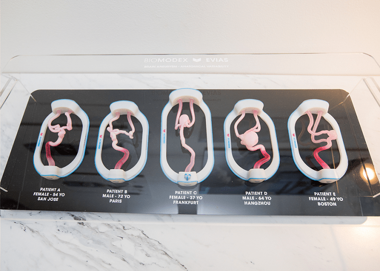
Biomodex’s 3D training models were created based on each patient’s unique CT angiography scans and printed using a Stratasys J750 Digital Anatomy 3D Printer. Biomodex Invivotech material was used to simulate the biomechanics of the aneurysm and the surrounding tissue. The models are made with various flexible photopolymers which, unlike single material models like silicone, allow control of the pliability of the target area, providing critical tactile feedback. The models are connected to the EVIAS station which includes a hydraulic system to replicate blood flow. The research study, led by RADIS lab’s clinical research technologist, Nicole Cancelliere (also lead co-author of the study), found the models to be a reliable and accurate reproduction of the aneurysm features and the parent vessel anatomy.
“The rehearsals allowed the operator to address issues, such as device sizing, and practice challenging manoeuvres, such as how to open flow diverter stents,” notes Cancelliere.


